

Red Flag Symptoms “Flashes, Floaters, Curtains”
Title: Red Flag Symptoms “Flashes, Floaters, Curtains”
Author: David F. Skanchy, MSIV, McGovern Medical School at UT Houston
Date: 09/20/18
Photographer: James Gilman, CRA, FOPS
Floaters are dark specks that float around inside the eye and may disrupt the field of vision. They may appear as spots, circles, cobwebs, clouds, or squiggly lines and are often easier to see while looking at a plain background or white computer screen. These floaters may drift with eye movement and dart away when attempting to look right at them. Floaters may be accompanied with flashes of light, often compared to streaks of lightning or camera flashes.
Causes:
The main cause of floaters and flashes is age-related changes of the vitreous humour (gel-like substance) that fills the eye. With time, the vitreous and collagen fibers inside the eye undergo degenerative changes forming clumps and strands. As light hits these strands in front of the retina, they may cast shadows onto the retina, causing the sensation described as floaters.
The vitreous may also begin to liquefy and collapse with age, pulling away from the retinal wall. As this vitreous shrinks it pulls toward the center of the eye causing traction on the retina. This force stimulates the retina causing the perception of flashing lights. Once the gel has completely detached, these flashes of light will subside.
Warning Signs:
Although floaters are usually benign, there are certain symptoms or situations that put patients at a greater risk of retinal tears or retinal detachments and would prompt urgent evaluation from an ophthalmologist. These include:
- New onset of a large floater or sudden increase in many floaters
- Sudden flashes of light, especially when persistent
- When a dark curtain or dark shadows appear in the visual field
- Floaters associated with blurred vision
- Recent eye trauma
- Floaters associated with high nearsightedness (high myopia)
Retinal Detachment
The retina is a thin layer of neural tissue that lines the inside wall of the eye. As light passes through the pupil and is focused onto the retina, it is converted into an electrical signal (phototransduction) that is carried by the optic nerve to the brain. The retina is like film in a camera that captures light and sends it to the brain, allowing us to visually process the world around us.
When the retina is separated from the retinal pigment epithelium (RPE) layer and choroid plexus below, it is cut off from part of its blood supply and can become ischemic. The macula, or central retina, is highly susceptible to damage when detached. There are three types of retinal detachments:
- Rhegmatogenous (most common): A tear in the retina allows vitreous fluid to enter and separate the retina from the underlying structures. A common cause of a retinal tear is posterior vitreous detachment (PVD), which is due to the vitreous shrinking and pulling away from the retinal wall as discussed earlier.
- Tractional: Scar tissue or other tissue pulls the retina away from underlying layers. This may happen in patients with severe diabetic retinopathy.
- Exudative: Fluid builds underneath the retina pushing it away from the underlying layers. This may be found in various inflammatory disease, kidney diseases, tumors, and with severely high blood pressure.
While a PVD is the most common benign cause of flashes and floaters, retinal detachments are the most worrisome and require prompt treatment to preserve vision. Other causes of flashes or floaters may include vitreous hemorrhage, posterior uveitis, oculodigital stimulation, and migraines.
Figures:
Figure 1 – What some floaters may look like from the perspective of the patient.

Figure 2 – Floaters can cast shadows onto the retina.
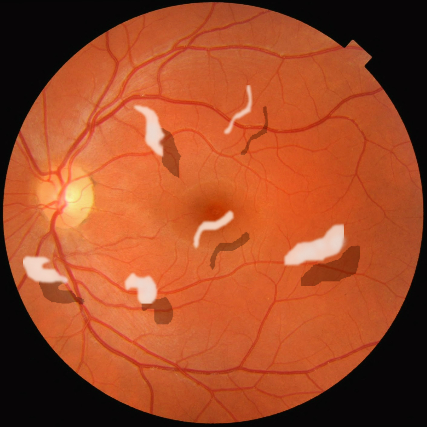
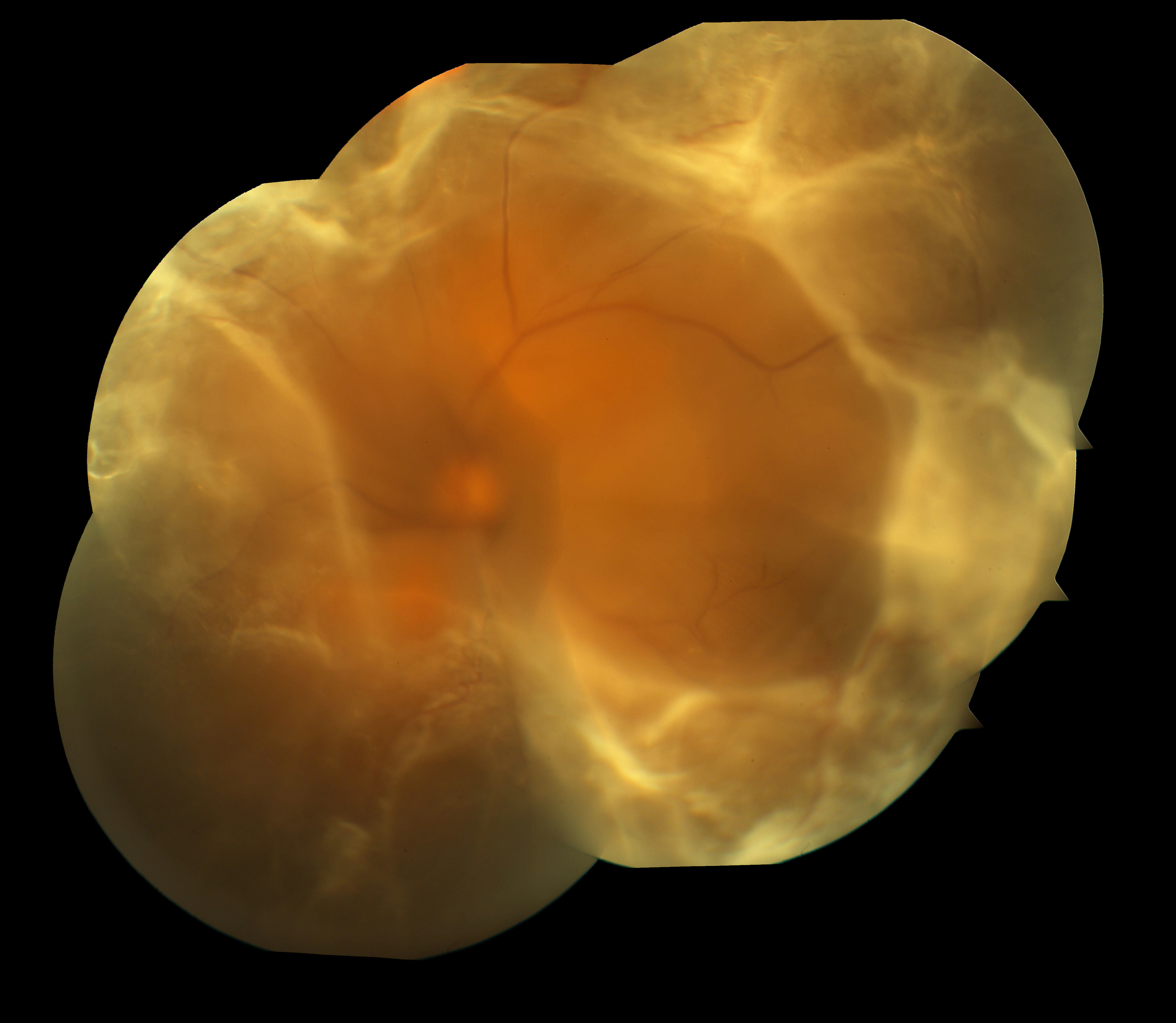
Figure 3/4 – OPTOS and color fundus photos of retinal detachments.

Faculty Approval: Griffin Jardine, MD
References:
Sharma P, Sridhar J, Mehta S. Flashes and floaters. Primary Care: Clinics in Office Practice. 2015 Sep 1;42(3):425-35.
Root, Timothy. OphthoBook. Beginner’s guide to the Retina. https://timroot.com/retina/ Accessed 20 September 2018.
Identifier: Moran_CORE_26803
Copyright statement: Copyright 2019. Please see terms of use page for more information