
Bull’s Eye Maculopathy as a Phenotypic Presentation of Stargardt Disease
Home / Retina and Vitreous / Hereditary Retinal and Choroidal Dystrophies
Title: Bull’s Eye Maculopathy as a Phenotypic Presentation of Stargardt Disease
Author: Troy Teeples, MSIV, University of Utah School of Medicine
Photographer: Unknown
Date: 8/14/2017
Image or video:

Image 1: Color fundoscopy shows pigment changes of the macula along with temporal pallor of the optic disc.

Image 2: Fundus Autoflourescence demonstrations central macular hypoautoflourescence with a surrounding hyperautoflourescent ring.
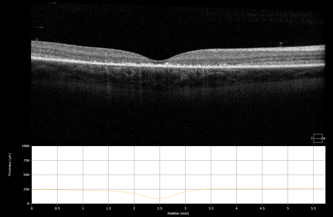
Figure 3: OCT demonstrations a discontinuous ELM layer, IS-OS dropout, and RPE disruption.
Keywords/Main Subjects:
Diagnosis: Stargardt Disease
Description of Image:
Patient Presentation: The patient is a previously healthy 7-year-old boy with no past medical history or family history of ocular disease who originally presented with blurry vision, problems with depth perception and central vision loss. In clinic he was found to have a visual acuity of 20/150 OD, 20/125 OS that was not corrected with refraction along with decreased color vision (Ishihara plates 4/8 OD, 5/8 OS). On fundoscopy he was found to have a Bull’s Eye Maculopathy OU with temporal pallor of the optic disc. No round pisciform flecks were seen on fundoscopy.
Stargardt Disease is the most common inherited macular dystrophy, though it may have many different phenotypic presentations. It is caused by accumulation of lipofuscin in the RPE, resulting from a mutation of the ABCA4 gene. With a faulty ABCA4 gene, the retina is not able to clear a toxic byproduct, bisretinoid-A2E, after the retinoid visual cycle, eventually resulting in RPE atrophy and photoreceptor cell death.
Phenotype Variation: Due to the large size of the ACBA4 gene (6.8kb), there are a significant number of disease-causing variants. In fact, over 500 distinct disease-associated mutations have been discovered that can cause Stargardt disease in addition to other ABCA4 associated diseases such as Cone Dystrophy, AMD, and Retinitis Pigmentosa. This patient presented with a 2 bp deletion resulting in a frameshift mutation on one allele as well as a missense mutation on the other, causing a Bull’s Eye Maculopathy as the phenotypic presentation. While some genotype-phenotype associations have been discovered, many remain yet to be determined. A prospective epidemiological study was performed over 12 months by Cornish and Ho in 2017 in order to determine the incidence of Stargardt Disease in the UK. Phenotypic presentations of the macula were quantified and of the 71 cases used in the study, 4% were found to have no macular abnormalities, 29% were found to have pigment mottling of the macula, 24% had a Bull’s Eye Maculopathy, 37% demonstrated macular atrophy and 36% had round pisciform flecks in the macula.
Imaging: Fundus Autoflouresence revealed a hypoautoflourescent macula with a surrounding hyperautoflourescent ring. An OCT revealed macular thinning, a discontinuous, ‘ratty’ ELM layer, IS-OS dropout, and disruption of the RPE.
References:
- http://eyewiki.aao.org/Stargardt_disease/Fundus_flavimaculatus
- Kurt Spiteri Cornish, Jason Ho, Susan Downes, Neil W. Scott, James Bainbridge, Noemi Lois. The Epidemiology of Stargardt Disease in the United Kingdom. Ophthalmology Retina,Volume 1, Issue 6, 2017, Pages 508-513., ISSN 2468-6530, https://doi.org/10.1016/j.oret.2017.03.001 (http://www.sciencedirect.com/science/article/pii/S2468653017300593)
Faculty Approval by: Griffin Jardine, MD
Footer:
- Copyright statement: Copyright Author Name, ©2016. For further information regarding the rights to this collection, please visit: URL to copyright information page on Moran CORE
- Attribution/citation suggestions
Disclosure (Financial or other): None