
Limbal Stem Cell Deficiency Image Report
Home / External Disease and Cornea / Clinical Approach to Depositions and Degenerations of the Conjunctiva, Cornea, and Sclera
Title: Limbal Stem Cell Deficiency
Author: Alex Wright, BS
Date: 07/25/2017
Images:
OS: 

OD: 
Keywords/Main Subjects: Limbal Stem Cell Deficiency
Diagnosis: Limbal Stem Cell Deficiency
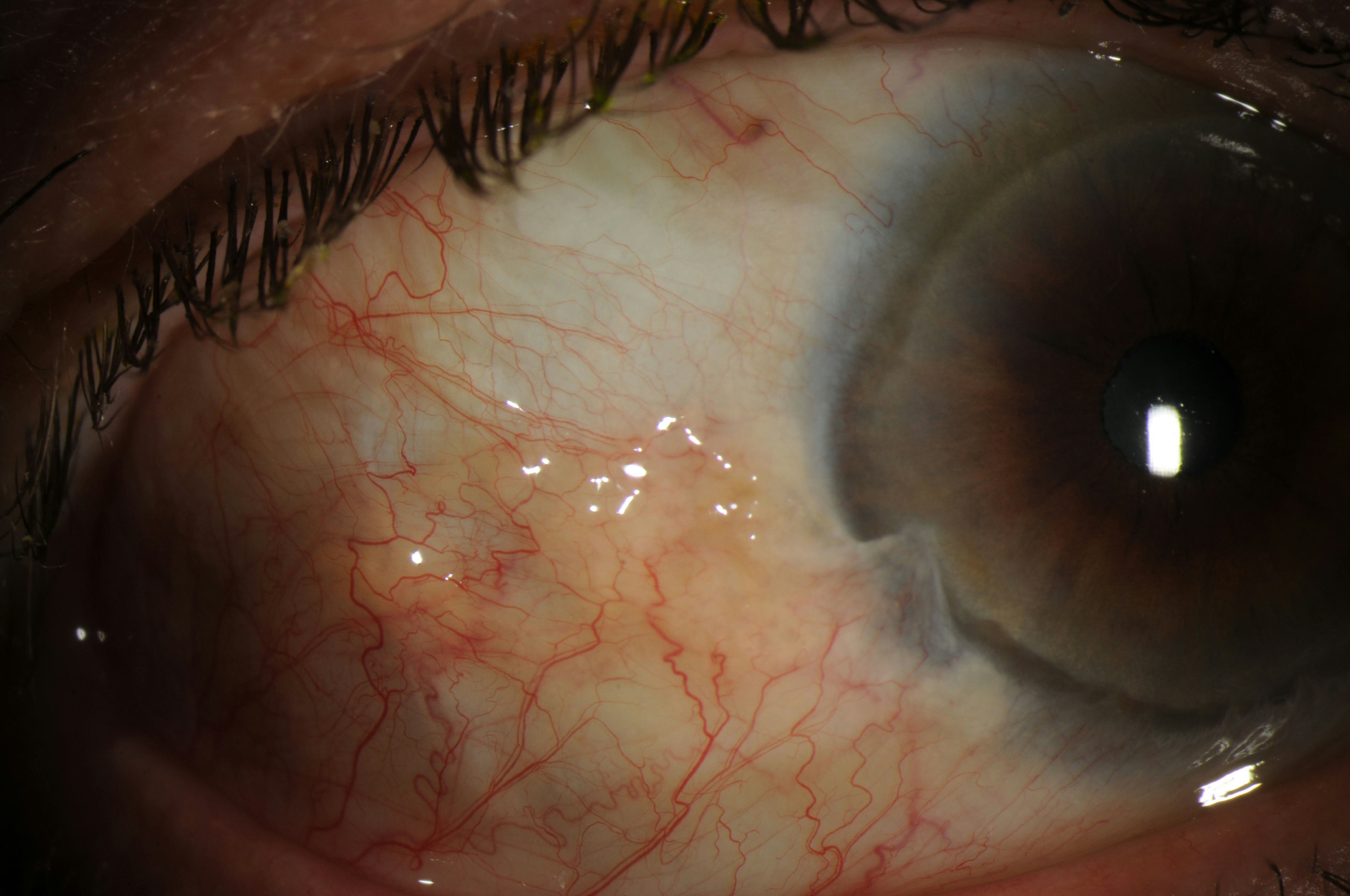
Description of Image: Limbal Stem Cell Deficiency (LSCD) occurs when the regenerative capacity of the corneal stem cells is impaired or damaged. The Palisades of Vogt, papilla-like structures of the limbus, are densely packed with basal cells, 10-15% of which are thought to be corneal stem cells. When these cells are damaged, the regenerative capacity of the cornea is gone. Thoft and Friend in 1983 originally hypothesized cell division in limbus plus further division of transit amplifying cells during their migration superficially and vertically equaled the amount of mature corneal stem cells lost and sluffed off in the tear film. As the cells are lost, the corneal surface begins to breakdown, which leads to further inflammation, conjunctival ingrowth, and subsequent neovascularization. Clinical symptoms include: photophobia, pain, conjunctivalization, corneal neovascularization, and recurrent ulceration. A diagnosis is established with clinical signs and symptoms and can be confirmed in some cases with impression cytology. Impression cytology looks for the presence of goblet cells on the cornea and helps identify conjunctivalization of the cornea, which is regarded as the most reliable diagnostic sign.
Pterygia can cause LSCD, but also must be differentiated from LSCD. Pterygia are often secondary to sun exposure and occur most commonly nasally. Pterygia are also more likely to be identified in younger patients. LSCD with pterygia are more likely bilateral and located in areas not exposed to the sun as often (notably inferiorly and superiorly). LSCD also occurs more commonly over the age of 50.
Etiologies of LSCD include: idiopathic, chemical/thermal burns, iatrogenic, autoimmune, and congenital/hereditary. Management initially if mild is aimed at controlling inflammation and the progression of the disease with topical steroids and topical cyclosporine drops. In severe cases surgery is often necessary to restore normal corneal epithelium. Many surgical procedures have been identified and studied, including: Conjunctival Limbal Autograft, Simple Limbal Epithelial Transplantation, Cultured Limbal Epithelial Transplantation, Living-Related Conjunctival Limbal Allograft, and Keratolimbal Allograft. When possible an autograft is preferred since allografts require systemic immunosuppression.
These images were taken from a 74-year-old Caucasian male with LSCD bilaterally. These images show a pterygium over the nasal cornea bilaterally and over the temporal cornea on the left eye. There is also signs of conjunctivalization and neovascularization inferiorly bilaterally.
References:
- Dua H S, Saini J S, Azuara-Blanco A, Gupta P. Limbal stem cell deficiency : Concept, aetiology, clinical presentation, diagnosis and management. Indian J Ophthalmol 2000;48:83-92
- Osei-Bempong C, Figuieredo FC, Lako M. The limbal epithelium of the eye – A review of limbal stem cell biology, disease and treatment. Bioessays 2012;35:211-219.
- Hatch KM, Dana R. The Structure and Function of the Limbal Stem Cell and the Disease States Associated With Limbal Stem Cell Deficiency. Int Ophthalmology Clin 2009;49:43-52
- Thoft RA, Friend J. 1983. The X, Y, Z hypothesis of corneal epithelial maintenance. Invest Ophthalmol Vis Sci 24: 1442–3.
- Kim KW, Mian SI. Diagnosis of Corneal Limbal Stem Cell Deficiency. CO-Ophthalmology 2017;28(4):355-362.
- Vadrevu VL, Fullard RJ. Enhancements to the conjunctival impression cytology technique and examples of applications in a clinico-biochemical study of dry eye. CLAO J 1994; 20:59–63.
- Holland EJ. Management of Limbal Stem Cell Deficiency: A Historical Perspective, Past, Present, and Future. Cornea 2015;34:S9-15.
Faculty Approval by: Dr. Brian Zaugg
Identifier: Moran_CORE_24246
Copyright statement: Copyright ©2017. Please see the Moran CORE terms of use for more information.
Disclosure (Financial or other): None