
Asymmetric Ptosis & Hering’s Law
Home / Orbit, Eyelids, and Lacrimal System
Contributors: BCK Patel, MD, FRCS; Mr. Raman Malhotra, FRCS
Photographer: BCK Patel MD, FRCS
Posted November 24, 2021
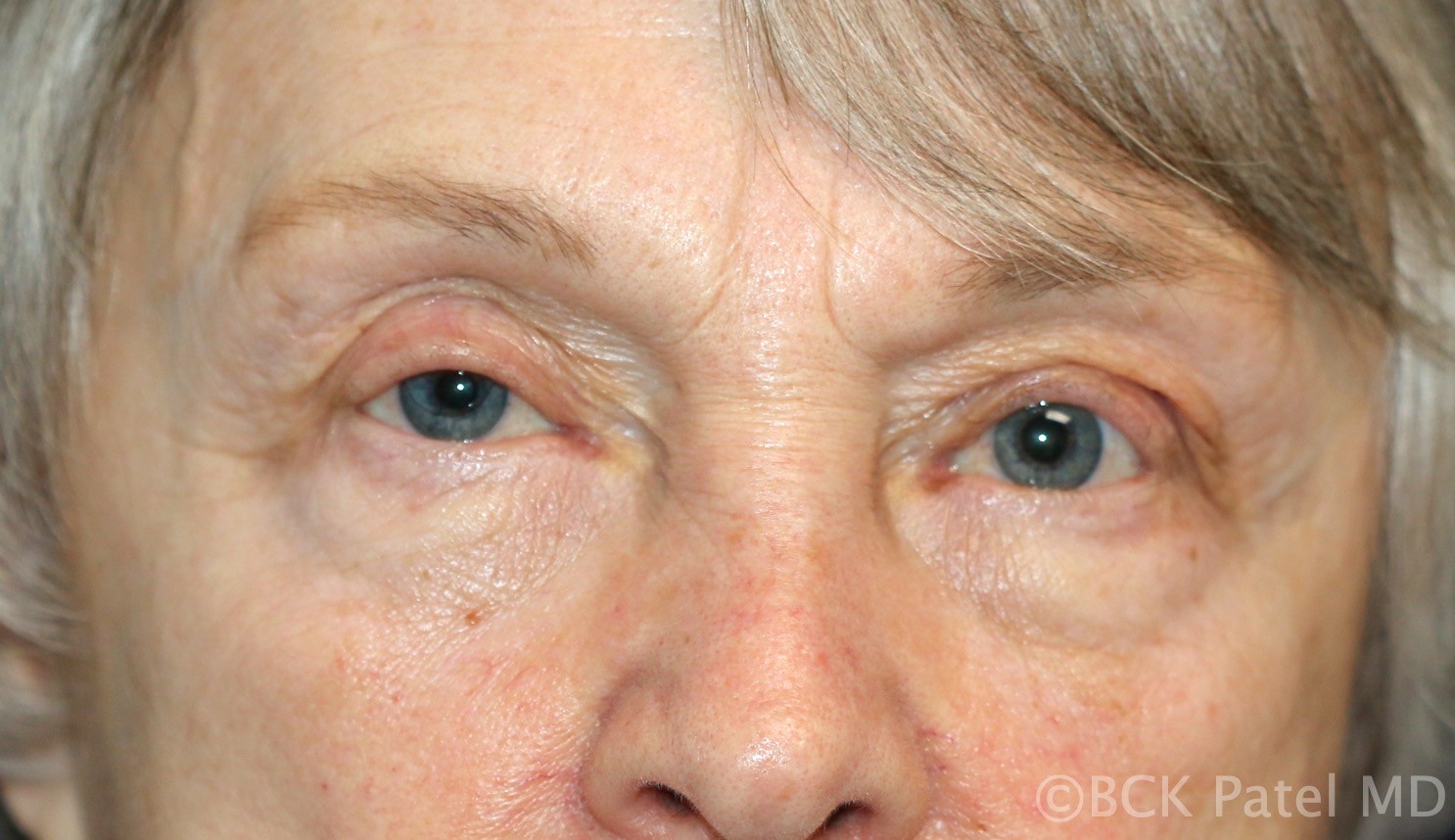
A 67-year-old female presents with a complaint of “my droopy left upper eyelid is interfering with my vision”. She notes that the left upper eyelid became droopy gradually over years. She has not had any intraocular surgery but has worn gas-permeable contact lenses for more than 20 years. She does not complain of any drooping of the right upper eyelid and she does not feel the right side interferes with her vision.

The patient has no history of thyroid disease or any other neuromuscular diseases. There is no history of trauma. She notes that the left upper eyelid “is much worse when I am tires and at the end of the day”.
Clinical examination shows a corneal reflex-eyelid margin distance of less than zero on the left side and 1 mm on the right. She has a high and poorly formed left upper eyelid skin crease with a deep superior sulcus. Her levator function is normal at more than 12 mm on both side. The right side shows a slightly raised upper eyelid skin crease which is more defined and there is early hollowing of the superior sulcus. Her left brow is higher than the right brow by 4 mm. Note the subtle prominence of the left medial fat pad which becomes revealed as the medial levator aponeurosis becomes “lateralized” with disinsertion. The orbital septum is weakest medially and this allows the medial fat pad to become more prominent. This is seen in many involutional ptosis cases. The lateralization of the levator aponeurosis also caused medial flattening of the upper eyelid as is obvious here. These are subtle signs that should be noted preoperatively and corrected surgically.
The common questions a surgeon may be faced with when encountering such a patient are as follows:
- If I have a droopy eyelid on one side, should I have surgery on one side or both sides?
- If I have surgery on one side because the Herring’s test did not show my “good” eyelid coming down, is there still a chance that the lid would become lower after one-sided surgery?
- What is the best option for me?
- When should one-sided droop of an eyelid be repaired? Should I wait until both sides droop?
Hering’s law of equal innervation states that eye muscles or eyelid muscles of each eye are equally innervated. Therefore, in the presence of a marked ptosis on one side, the equal innervation will result in an artificially elevated opposite eyelid. If the ptotic eyelid is lifted, the opposite eyelid will usually fall because the stimulus that lifts the ptotic eyelid is lifted and the same will apply to the opposite side.
Q 1. If I have a droopy eyelid on one side, should I have surgery on one side or both sides?
- 1. When the Hering’s is positive, it makes sense to operate upon both upper eyelids at the same time. It should be remembered that when the Hering’s test is done (by lifting the droopy lid and watching the “normal” eyelid for drooping), the relaxation of the “normal” side may not always be obvious or dramatic. Subtle changes can be easily missed. So a careful, and, if necessary, a repeat examination is vital to help make a decision.
Q 2. If I have surgery on one side because the Hering’s test did not show my “good” eyelid coming down, is there still a chance that the lid would become lower after one-sided surgery?
A 2. Absolutely. The preoperative Hering’s law test is usually predictive, but there can be a mild ptosis on the unaffected side which can become manifest after unilateral surgery. Swelling, bruising, etc after surgery will make the operated eyelid look persistently droopy for some days (even weeks) and therefore, it is important to give the body a chance to heal and the neurological responses (Hering’s in other words) to manifest themselves. We will generally not make any judgement about the final lid height for at least two months after surgery, sometimes longer.
Q3. What is the best option for me?
A3. How many different shades of grey are there? Horses for courses. One should not just assess the eyelids. The relationship of the eyelids to the brows (depending upon racial characteristics, sun exposure, contact lens wear, age, etc) has to be considered. Asymmetry of the face (we all have asymmetric faces) becomes slowly-but-surely more apparent as we age and this applies to the periorbital area and eyelids as well. People forget to look at the position of the lower eyelids when addressing a droopy eyelid or eyelids. We will all have one lower lid a little different (lower, fuller, etc) when compared to the other. So, after an assessment of all these factors, and with the help of a full-face photograph, options can be discussed.
Q 4. When should one-sided droop of an eyelid be repaired? Should I wait until both sides droop?
This is simple. If a slightly droopy eyelid does not bother you functionally or cosmetically, leave it well alone. Mild and even moderate asymmetry of the face and the eyelids is common. If you choose to have surgery on the droopy upper eyelid only, be prepared to see asymmetry after surgery with the operated side looking higher than the unoperated side.
This patient underwent an anterior approach left upper eyelid ptosis repair. The orbital septum was opened and the fat released but no fat was removed. The fat was advanced to fill the deep superior sulcus and attached to the orbicularis to provide a fullness above the eyelid skin crease. The eyelid skin crease was recreated with imbrication sutures which picked up the levator aponeurosis in the eyelid skin closure.
The patients’ appearance two months after surgery was as follows:
Note the following:
- The left brow that was higher than the right brow has relaxed and is about the same height as the right brow.
- The left ptosis is corrected with an improvement in the skin crease and an improvement in the left deep superior sulcus.
- The right upper eyelid now looks ptotic. However, the corneal reflex-lid margin distance is only slightly smaller.
- Note that the right superior sulcus is now deeper and the skin crease appears much higher. With Hering’s law, when the stimulus to keep the opposite eyelid up is removed, the levator will relax and any underlying levator disinsertion may become manifest with a difference in the skin crease (which will be higher) and the sulcus (which will be deeper as the disinserted levator and the attached septum pull back revealing the deeper superior sulcus. It is important to remember that this type of apparent deepening of the superior sulcus with Hering’s law is frequently missed and the patient my wrongly assume that the surgeon caused this quite dramatic difference.
- Note the correction of the prominent medial fat pad on the left and also the correction of the medial flattening of the upper eyelid.
- By recreating the skin crease, we can also evert the ptotic eyelashes as was done here.
This particular patient was still not bothered visually by the right upper eyelid so she elected not to have any surgery on the right side. However, other patients may choose to have the now apparent droop of the right upper eyelid corrected. The surgeon should aim to give the patient a symmetrical skin crease, and fill the superior hollow (sulcus) to achieve as much symmetry as possible. This surgery may be performed at any time the patient wishes.

References
- Gay AJ, Salmon ML, Windsor CE. Hering’s law, the levators, and their relationship in disease states. Arch Ophthalmol 1967;77(2):157-160 https://pubmed.ncbi.nlm.nih.gov/6019006/
- Koka K, Patel BCK. Ptosis Correction. StatPearls, Treasure Island (FL): StatPearls Publishing. July 2021. https://pubmed.ncbi.nlm.nih.gov/30969650/
- Wong MB, Maamari RN, Couch SM. Contralateral eyelid elevation following unilateral upper eyelid retraction repair. Orbit 2021 May 25:1-5 https://pubmed.ncbi.nlm.nih.gov/34030599/
Identifier: Moran_CORE_119265
Copyright statement: Copyright 2021. Please see terms of use page for more information