
Preseptal vs Orbital Cellulitis
Home / Basic Ophthalmology Review / Orbit
Name: Paul D Chamberlain, 4th year medical student, Baylor College of Medicine; Reese Feist, Chief Resident, University of Utah Moran Eye Center.
Topic: Preseptal vs Orbital Cellulitis
Terminology and Anatomy
Differentiating orbital from preseptal cellulitis is extraordinarily important given that orbital cellulitis has the potential to cause a compartment syndrome within the eye socket resulting in irreversible vision loss to the affected eye. The orbital septum is a membranous sheath extending from the periosteum of the orbit to the tarsal plate located in the eyelid, and is the key anatomical structure in differentiating preseptal from orbital cellulitis. The orbit (eye socket) is the bony structure in which the globe (eyeball) is housed, and it also contains extraocular muscles, fat, and the blood vessels and nerves that supply these structures. Orbital cellulitis (Image 1), also called post-septal cellulitis, is inflammation of the soft tissues (muscles, fat, and connective tissue) of the orbit most commonly from infection. It is important to remember that in orbital cellulitis, the globe itself is not infected or inflamed. Because the orbit is surrounded by the frontal, ethmoid, and maxillary sinuses, infection often results from extension of a sinus infection.
In comparison, pre-septal cellulitis (Image 2), also known as peri-orbital cellulitis, is an infection of the eyelids and surrounding soft tissues that are anterior to the orbital septum. Both orbital cellulitis and preseptal cellulitis are more common in children, and preseptal cellulitis is much more common that orbital cellulitis.

A patient with orbital cellulitis. Note: orbital cellulitis can take on a variety of clinical manifestations and this image should not be taken as a gold standard to which an examiner compares a patient.

A patient with preseptal cellulitis.
Clinical Manifestations
A number of signs may alert the examiner to the presence of orbital cellulitis (table 1). Patients typicall present with erythema and edema of the eyelids. In orbital cellulitis, they erythema and edema can abruptly stop at the arcus marginalis, where the orbital septum inserts into the periosteum. Preseptal cellulitis typically expands beyond this landmark. Patients may present with eye pain, especially with eye movements due to irritation of inflamed muscles. Partial or complete ophthalmoplegia (inability to move the eye in one or more directions) and associated diplopia (double vision) due to inflammation of extraocular muscles or associated cranial nerves. In orbital cellulitis, inflammation and/or an orbital abscess can displace the globe, often pushing it forward or outward which is called proptosis. The presence of proptosis is a medical emergency to evaluate for compartment syndrome. The eyelids can be swollen and in severe cases swollen shut. Eyelids being swollen shut can be present in either orbital or preseptal cellulitis, and is not as helpful in distinguishing between the two. Visual acuity may be decreased but is often unaffected, and a normal visual acuity should not rule out orbital cellulitis. There is often a history or sinusitis or dental abscess. However, there may be no clear source of inflammation, and it may not even be infectious, as with idiopathic orbital inflammatory syndrome.
Preseptal cellulitis is much more common than orbital cellulitis and also presents with eye pain and erythema of the eyelid and surrounding skin and soft tissue. The inflammation may be great enough to tightly close the eyelids as well (image 3), and they should be opened for a visual inspection. Pre-septal cellulitis does not cause loss of vision and if visual acuity is decreased it is likely because of a poor exam or a more severe infection. Preseptal cellulitis may also arise from sinusitis, but may also arise secondary to local trauma, foreign body, or bug bite (Image 4).

Preseptal cellulitis with mechanical ptosis or droopy eyelid due to edema and erythema of the eyelids. Note that there is no proptosis and the erythema doesn’t abruptly stop at the orbital rim, making it clinically less likely to be orbital cellulitis.
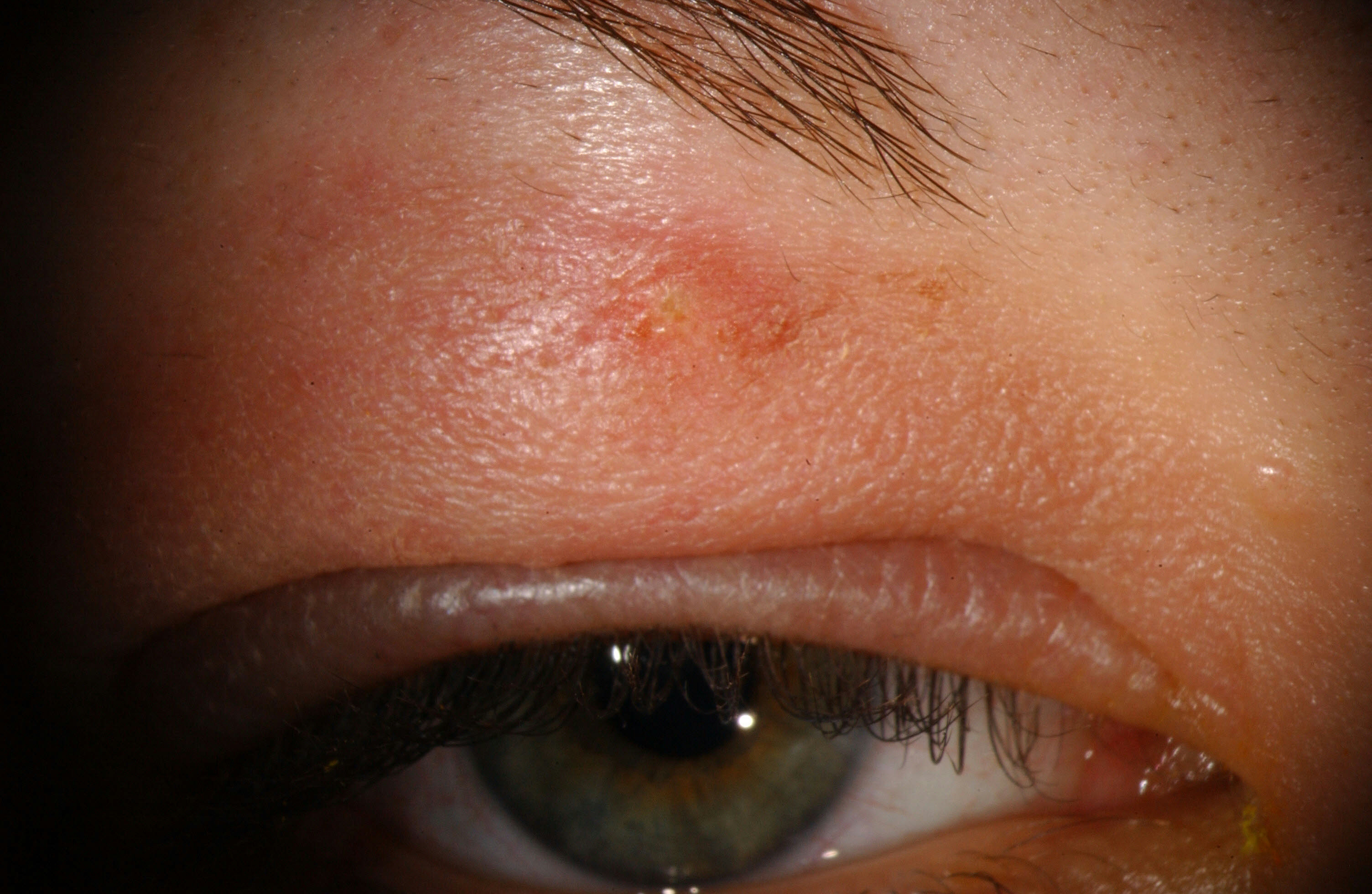
A patient with preseptal cellulitis secondary to a bug bite.

Orbital cellulitis. Note the bullous, edematous conjunctiva (conjunctival chemosis), proptosis and the delineation of swelling around the orbital rim. This patient underwent an urgent lateral canthotomy/cantholysis.
Table 1: Comparison of clinical, historical, and diagnostic characteristics of preseptal and orbital cellulitis.
| Characteristic | Preseptal Cellulitis | Orbital Cellulitis |
| Eye pain | May be present | Yes |
| Eyelid erythema and/or tenderness | Yes | Yes |
| Pain with eye movements | No | May be present |
| Ophthalmoplegia ± diplopia | No | May be present* |
| Proptosis | No | May be present* |
| Vision loss | No | May be present* |
| RAPD | No | May be present* |
| Fever | Usually not present | Usually present |
| Intraocular Pressure (IOP) | Normal | May be elevated* |
| Resistance to Retropulsion | None | Present* |
| History of sinusitis | May be present, but often not | Present more often than not |
| CT or MRI imaging | Shows inflammation only anterior to the orbital septum | Show post-septal involvement of the inflammation. |
| Blood Cultures | Very rarely has bacteremia | Bacteremia may be present |
| *Emergent signs and symptoms that might warrant immediate lateral canthotomy/cantholysis | ||
Work-up and Treatment
As previously discussed, the first step is to determine whether a patient has orbital cellulitis, which requires orbital imaging, admission, blood cultures and IV antibiotics. Begin with a thorough ophthalmologic exam, with particular attention to visual acuity, pupillary testing for a relative afferent pupillary defect (RAPD), intraocular pressure, and assessment of proptosis and eye motility. In cases of question, Computed tomography (CT) with and without contrast of the orbits and sinuses should be ordered to look for evidence of post-septal involvement. If a diagnosis of orbital cellulitis is made, the patient needs to be immediately assessed monitored for signs of compartment syndrome and optic neuropathy which would warrant an emergent lateral canthotomy/cantholysis. This procedure allows for anterior expansion of orbital contents which relieves pressure within the orbit in restores blood flow to those structures. Even after initiating antibiotics the swelling may increase for the first 24-48 hours, so frequent re-evaluation is warranted. Immediate surgery may be indicated if there is evidence of a subperiosteal abscess, orbital abscess, or extension of the infection into the cranium. Consider consulting an otolaryngologist for management of sinus disease.
Patients whose history and examination are consistent with preseptal cellulitis without symptoms of orbital cellulitis may be treated as an outpatient with oral antibiotics. Patients with preseptal cellulitis who are appropriately treated typically recover completely without any permanent sequelae. If treated appropriately, patients with orbital cellulitis also often have good outcomes. However, failure to diagnose and treat orbital cellulitis in a timely manner may result in permanent vision loss. An ophthalmologist should be consulted in all cases of orbital or preseptal cellulitis. However, assessing for vision threatening orbital cellulitis should not be postponed until an ophthalmologist is available as an irreversible ischemic optic neuropathy can occur in less than 90 minutes.
References:
Amin N, Syed I, Osborne S. Assessment and management of orbital cellulitis. British Journal of Hospital Medicine. 2016; 77(4):216-20.
Hauser A, Fogarasi S. Periorbital and orbital cellulitis. Pediatrics in Review. 2010; 31(6):242-9.
Meara DJ. Sinonasal disease and orbital cellulitis in children. Oral Maxillofacial Surgery Clinics of North America. 2012; 24(3):487-96.
Rashed F, Cannon A, Heaton PA, Paul SP. Diagnosis, management and treatment of orbital and periorbital cellulitis in children. Emergency Nurse. 2016; (24(1):30-5.
Identifier: Moran_CORE_24521