
Fundus photo and OCT of Best’s vitelliform macular dystrophy (BVMD)
Home / Pediatric Ophthalmology and Strabismus / Disorders of the Retina and Vitreous
Title: Fundus photo and OCT of Best’s vitelliform macular dystrophy (BVMD)
Author (s): Gavin Gorrell, 4th Year Medical Student, University of New Mexico School of Medicine
Date: 06/24/2017
Image:

Keywords/Main Subjects: Best’s Disease; Best’s vitelliform macular dystrophy (BVMD); bestrophinopathies
Secondary CORE Category: Home / Retina and Vitreous / Hereditary Retinal and Choroidal Dystrophies
Diagnosis: Best’s vitelliform macular dystrophy (BVMD)
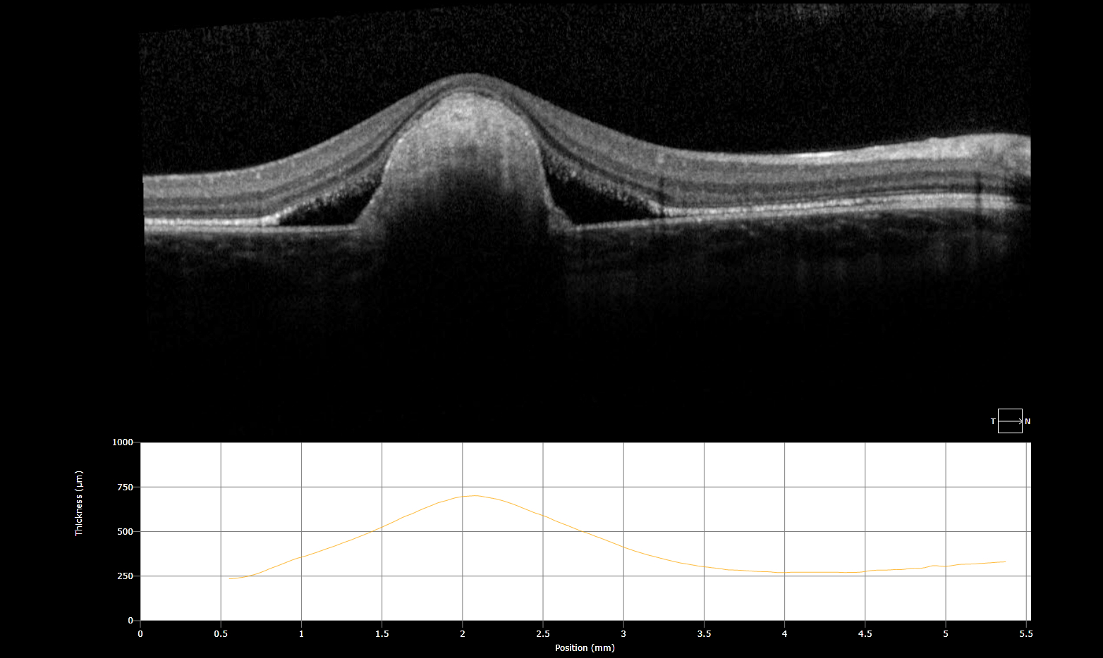
Description of Image:
- Egg-yolk appearance of lipofuscin accumulation at the central macula
- Subretinal fluid and lipofuscin accumulation causing RPE detachment
Pathogenesis (&Presentation): Best vitelliform macular dystrophy (BVMD) is the second most common hereditary macular dystrophy and is autosomal dominant with variable penetrance and expression. BVMD is the most common of the “bestrophinopathies”, a group of diseases which all contain various mutations to the BEST1 gene. BEST1 (previously termed VMD2), codes for bestrophin 1, a transmembrane protein in the RPE believed to be involved in anion transport and calcium signaling. Through unclear mechanisms, dysfunction of bestrophin 1 in BVMD leads to accumulation of lipofuscin (a retinal breakdown pigment) between Bruch’s membrane and RPE. This debris leads to the characteristic subretinal egg-yolk lesion.
Presentation: In early disease, usually beginning between ages 3 and 15, fundoscopy shows significant vitelliform (vitellus is latin for egg-yolk) lesions to the central macula, however patients may be asymptomatic or have only minimal loss in visual acuity. Over time the lipofuscin in the initial lesions disperses and is resorbed, leading to a “scrambled egg” appearance. A slow progression to RPE atrophy is expected with vision loss typically stabilizing around 20/30-20/200. Rapid decline in VA should cause concern for choroidal neovascularization, a feared complication that occurs in about 20% of patients (eyewiki).
Diagnosis & Differential:
Diagnosis is primarily made with family history and clinical appearance. Electro-oculography (EOG), which demonstrates abnormally low light/dark ratio in Best’s disease (<1.5), is often obtained to confirm the diagnosis. Genetic testing for BEST1 mutation is also available. Providers should consider OCT and/or fluorescein angiography to evaluate for choroidal neovascularization.
DDx: Adult-onset vitelliform macular dystrophy, toxoplasmosis retinochoroiditis, AMD, Bull’s eye maculopathy
Management: There is no current treatment available for Best’s disease, however patients should be monitored for choroidal neovascularization.
References:
Freund, K. Bailey, David Sarraf, William F. Mieler, and Lawrence A. Yannuzzi. “Hereditary Chorioretinal Dystrophies.” In The Retinal Atlas, 2nd ed., 13–231. Philadelphia, PA, 2017. https://www-clinicalkey-com.libproxy.unm.edu/#!/content/book/3-s2.0-B9780323287920000028?scrollTo=%23hl0000946.
Johnson, Adiv A., Karina E. Guziewicz, C. Justin Lee, Ravi C. Kalathur, Jose S. Pulido, Lihua Y. Marmorstein, and Alan D. Marmorstein. “Bestrophin 1 and Retinal Disease.” Progress in Retinal and Eye Research 58 (May 2017): 45–69. doi:10.1016/j.preteyeres.2017.01.006.
MacDonald, Ian M., and Thomas Lee. “Best Vitelliform Macular Dystrophy.” In GeneReviews(®), edited by Roberta A. Pagon, Margaret P. Adam, Holly H. Ardinger, Stephanie E. Wallace, Anne Amemiya, Lora JH Bean, Thomas D. Bird, et al. Seattle (WA): University of Washington, Seattle, 1993. http://www.ncbi.nlm.nih.gov/books/NBK1167/
“The Electroretinogram and Electro-Oculogram: Clinical Applications by Donnell J. Creel – Webvision.” Accessed June 24, 2017. http://webvision.med.utah.edu/book/electrophysiology/the-electroretinogram-clinical-applications/.
“Best Disease – EyeWiki.” Accessed June 24, 2017. http://eyewiki.aao.org/Best_Disease
Faculty Approval by: Griffin Jardine, MD
Disclosure (Financial or other): None
Copyright statement: Copyright 2016. Please see terms of use page for more information.