
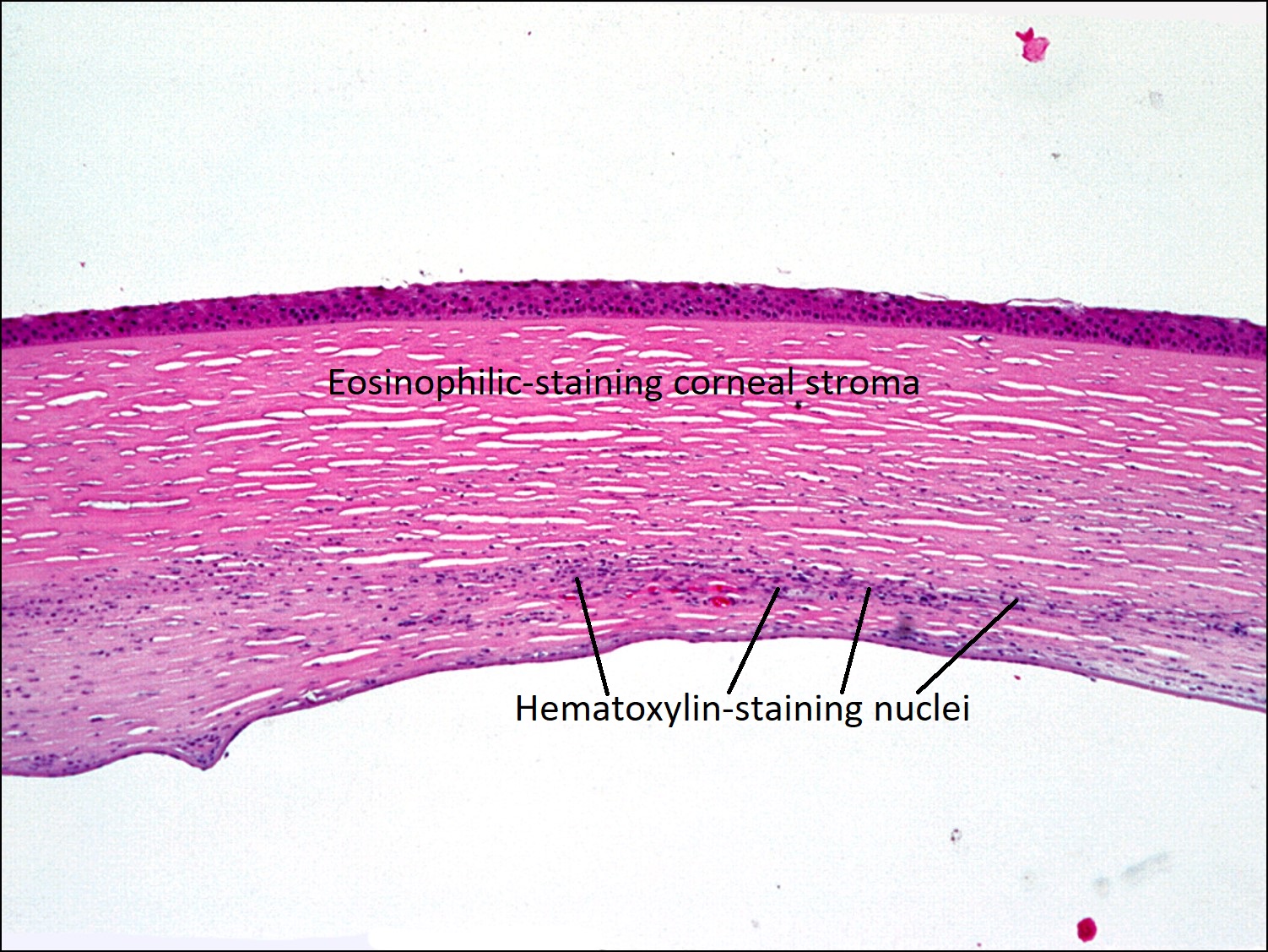
Stains and Processing
Home / Ophthalmic Pathology / Stains


| Stains | Colors | Examples |
|---|---|---|
| H&E | Blue: nucleus
Red/pink: cytoplasm |
Routine tissue stain |
| PAS | Magenta: basement membrane glycogen and proteoglycans | Descemet membrane |
| Alcian Blue | Blue: acid mucopolysaccharides | Macular corneal stromal dystrophy |
| Colloidal Iron | Blue: acid mucopolysaccharides
Red/purple: collagen |
Macular corneal stromal dystrophy |
| Alizarin Red | Red: calcium | Band keratopathy |
| Von Kossa | Black: calcium phosphate | Band keratopathy |
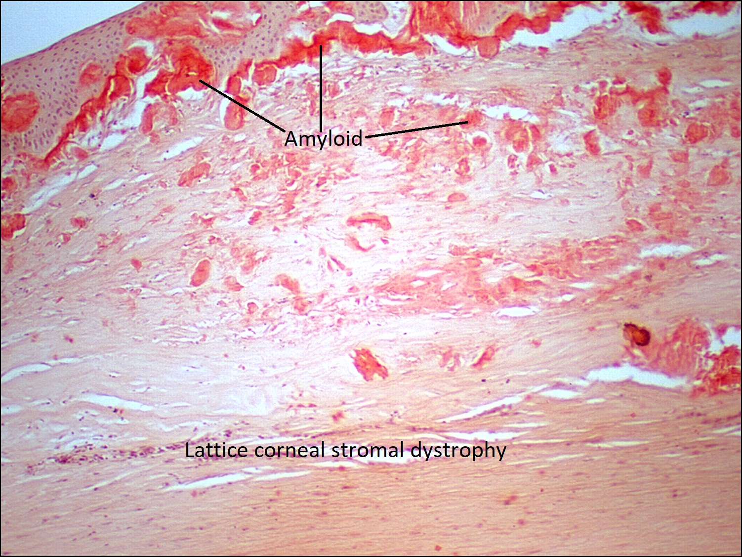
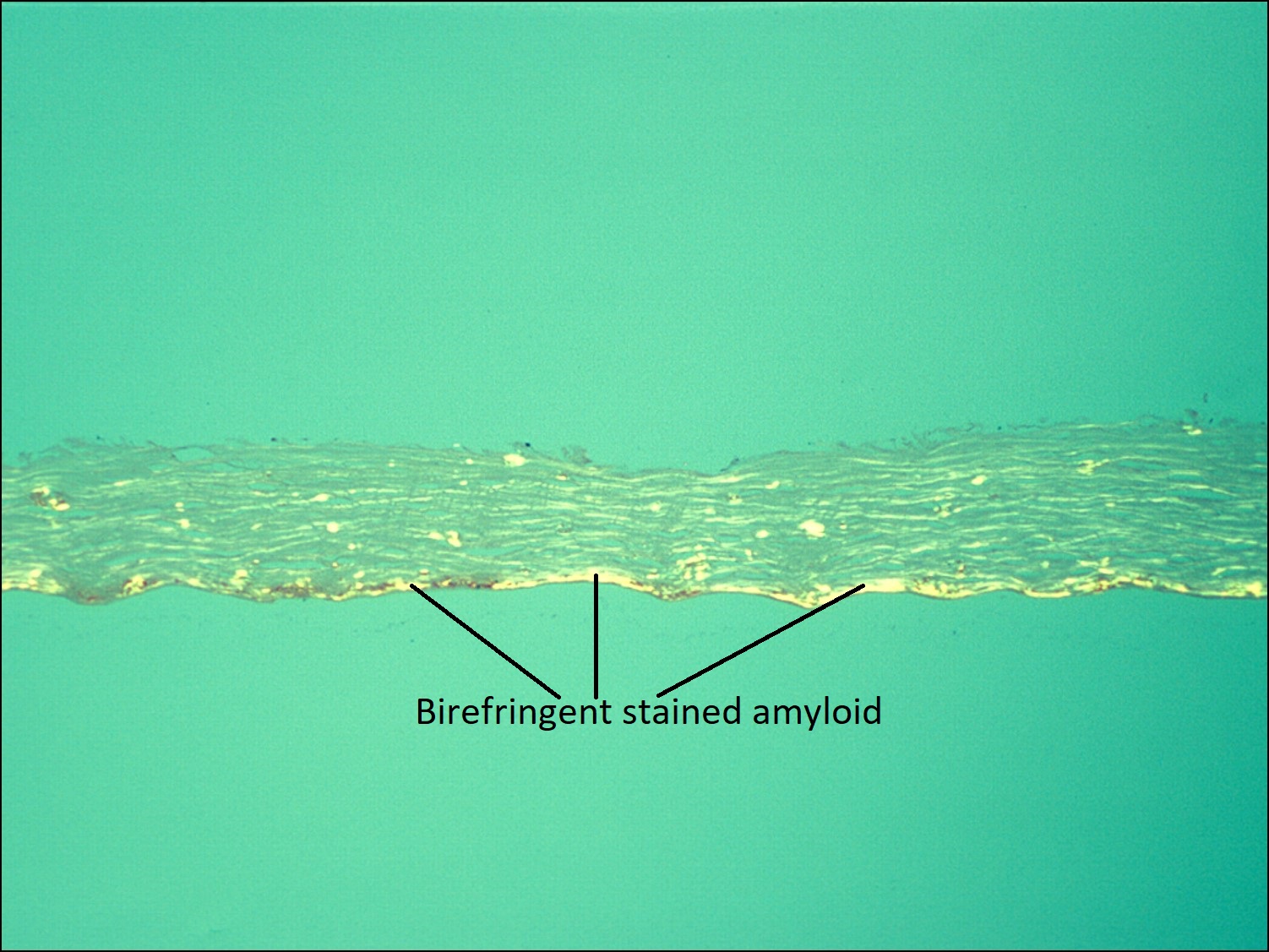
| Congo Red | Orange with red-green birefringence: amyloid | Lattice corneal stromal dystrophy |
| Crystal Violet | Purple/violet: amyloid | Lattice corneal stromal dystrophy |
| Thioflavin T | Fluorescent yellow: amyloid | Lattice corneal stromal dystrophy |
| Masson’s Trichrome | Blue: collagen
Dark red/purple: nuclei Red: cytoplasm, muscle |
Granular corneal stromal dystrophy |
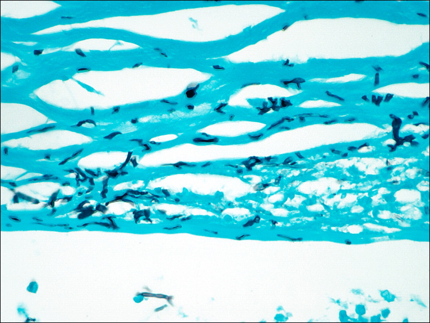
| Gram Stain | Blue: Gram-positive bacteria
Red: Gram-negative bacteria Yellow: corneal stroma |
Bacterial infections |
| Ziehl-Neelsen | Red: acid-fast organisms
Blue: background |
Tuberculosis |
| GMS | Black: fungal elements
Blue-green: background |
Fungal infections |
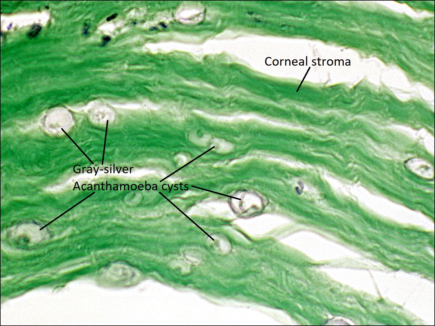
| Gridley | Gray-silver: acanthamoeba cysts
Green: background |
Acanthamoeba infections |
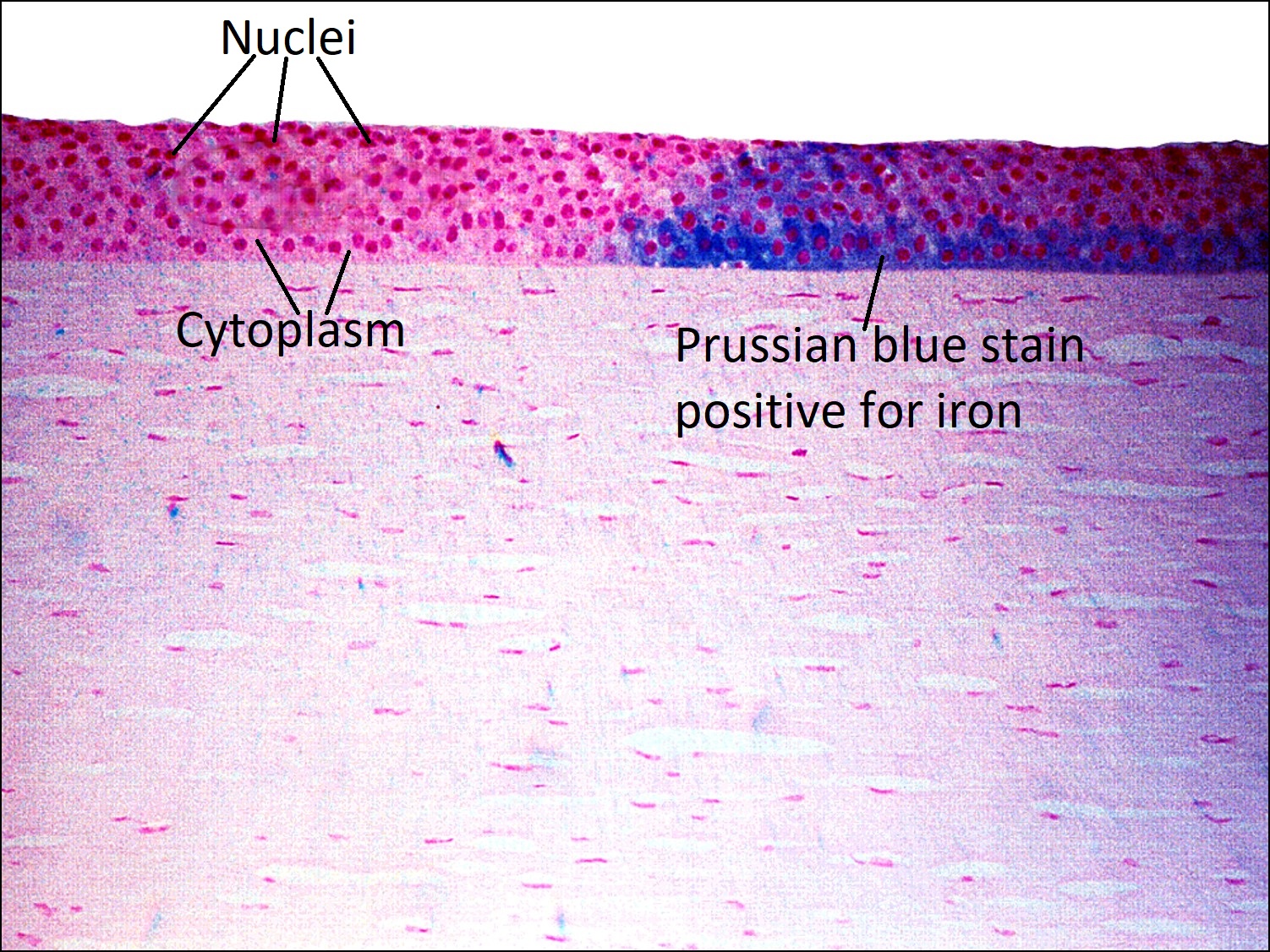
| Prussian Blue | Blue: iron | Hemosiderosis bulbi |
| Verhoeff-van Gieson | Black: elastic fibers | Temporal artery biopsy |
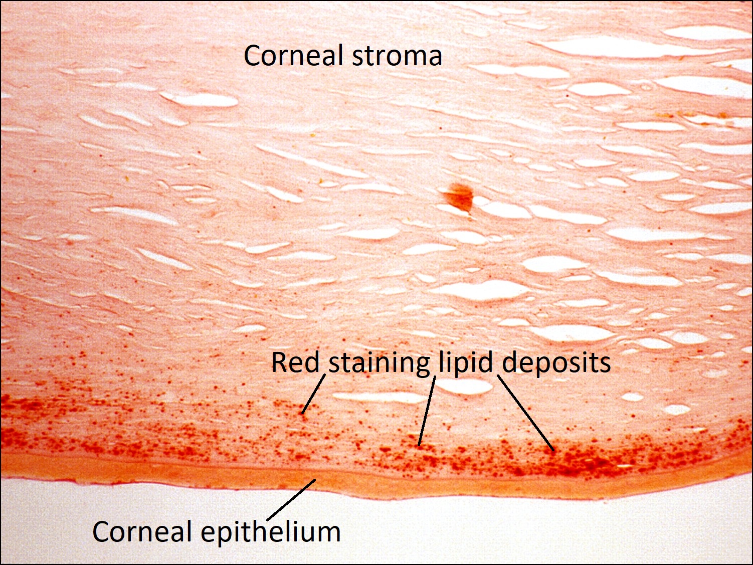
| Oil Red-O | Red: lipid | Sebaceous carcinoma |
| Sudan Black | Black: lipid | Sebaceous carcinoma |