
Multimodal imaging of choroidal metastases from adenoid cystic carcinoma of the submandibular gland.
Home / Ophthalmic Pathology / Ocular Involvement in Systemic Malignancies
Title: Multimodal imaging of choroidal metastases from adenoid cystic carcinoma of the submandibular gland.
Author: Brian M. Besch
Date: 06/26/2017
Images:

Keywords/Main Subjects: adenoid cystic carcinoma, choroidal metastasis, ophthalmic pathology, intraocular tumors
Diagnosis: adenoid cystic carcinoma of left submandibular gland with bilateral choroidal metastases
Description of Image: Adenoid cystic carcinoma (ACC) is a rare malignancy originating from multiple sites, but typically arises in the head and neck, and most commonly the salivary glands. Histologically, the lesion is composed of epithelial and myoepithelial cells in three configurations – tubular, cribiform, or solid; the latter is the most aggressive type. The malignancy generally arises between ages 40-60 and has an indolent, but nonetheless persistent course generally refractory to treatment. Initial management is wide surgical resection, typically followed by adjuvant external beam radiation or chemotherapy with cisplatin or melphalan. Distant metastases are common later in the disease course; one report indicates a mean of 48 months following initial management. Metastasis to the lung is most common. Choroidal metastases are rare; at the time of writing, the associated images represent the seventh reported case in the literature. While the limited reports of ACC in general suggest a slight female predominance, of the 6 cases involving choroidal metastases previously described, 5 were in women. All 5 primary tumors arose from submandibular glands, while the single male case derived from the hard palate.
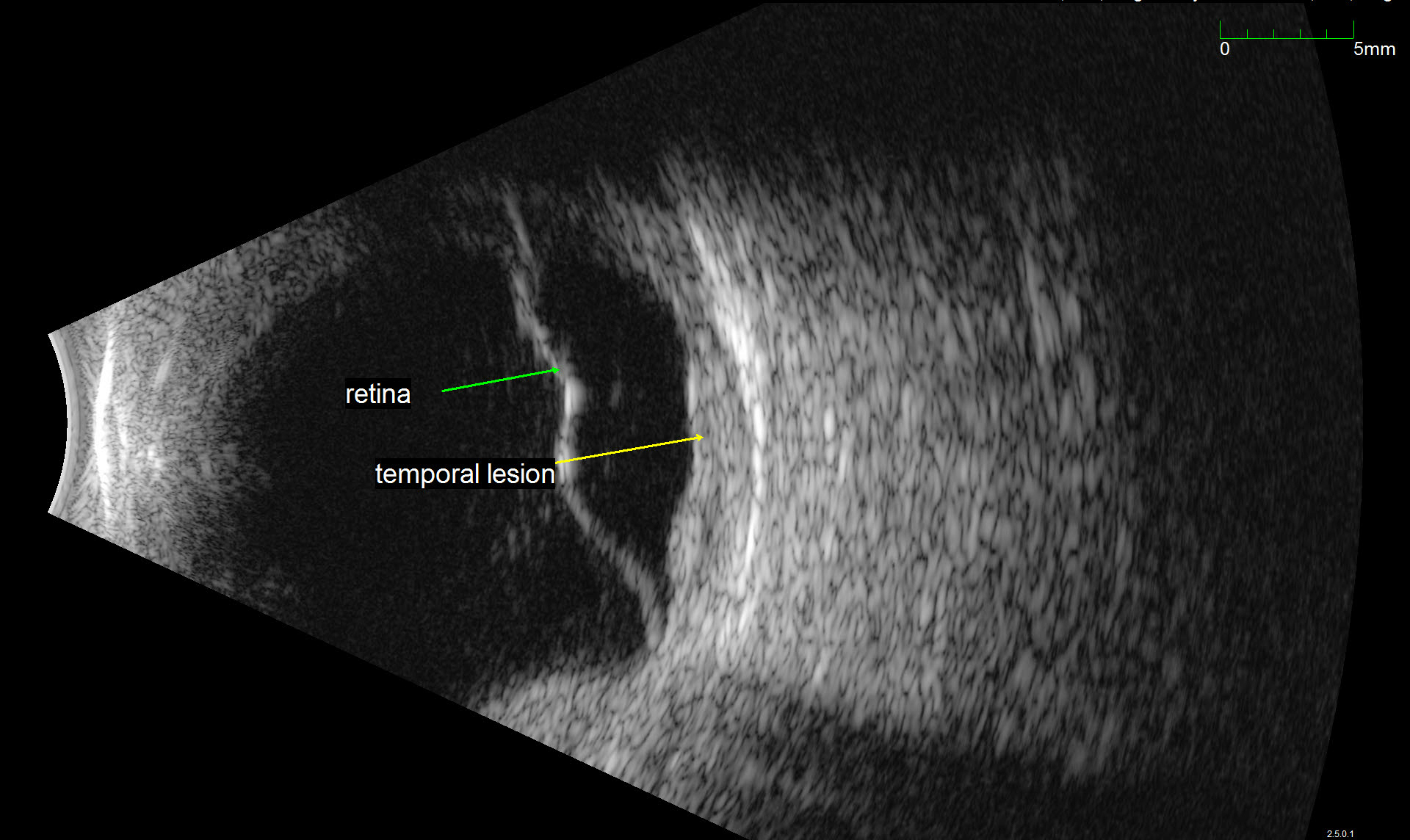
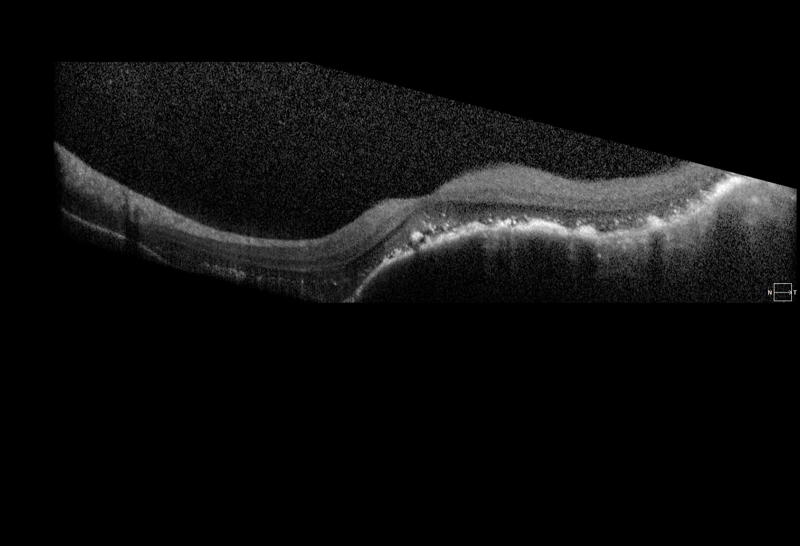
The images represent bilateral choroidal ACC metastases from a 57 year old female. The fundus montage illustrates two metastatic lesions in the left eye; one involves the macula, and is associated with inferior serous retinal detachment. A labeled ultrasound frame indicates the larger temporal lesion and retinal detachment. The OCT scan illustrates a large choroidal mass, marked retinal architecture distortion, and sub-retinal serous edema. The patient initially presented with a lump in the submandibular region and underwent FNA with inconclusive cytology. On re-evaluation and head/neck CT imaging, she was noted to have a submandibular mass and an enlarged lymph node; she subsequently underwent left submandibular gland excision and neck dissection followed by adjuvant chemoradiation. Post-surgical CT of the chest, abdomen, and pelvis revealed multiple small pulmonary lesions bilaterally concerning for metastases. Manifestation of the choroidal lesions was noted on fundus exam approximately two years following the initial diagnosis.
References:
Cai, Qian et al. “Adenoid Cystic Carcinoma of Submandibular Salivary Gland With Late Metastases to Lung and Choroid: A Case Report and Literature Review.” Journal of Oral and Maxillofacial Surgery 72.9 (2014): 1744–1755.
Shie, Jerry A. LDS et al. “Bilateral Choroidal Metastasis From Adenoid Cystic Carcinoma of the Submandibular Gland.” Retina 20.4 (2000): 406–407.
Faculty Approval by: Griffin Jardine, MD; Akbar Shakoor, MD
Identifier: Moran_CORE_24186
Copyright statement: Copyright 2017. Please see terms of use page for more information.