
How to Use the Direct Ophthalmoscope
Home / Basic Ophthalmology Review / Direct Ophthalmoscope
Title: How to Use the Direct Ophthalmoscope
Authors: Tania Padilla Conde, 4th Year Medical Student, University of South Dakota Sanford School of Medicine; Christopher Bair, MD and Michele Burrow, MD
Date: 08/10/2018
Videographer: Ethan Peterson
LOCATION: Medical Student Education Outline > I. Introduction to the Eye Exam > Direct Ophthalmoscope > Using a Direct Ophthalmoscope – VIDEO
Learning Objectives
- Understand the utility of the direct ophthalmoscope
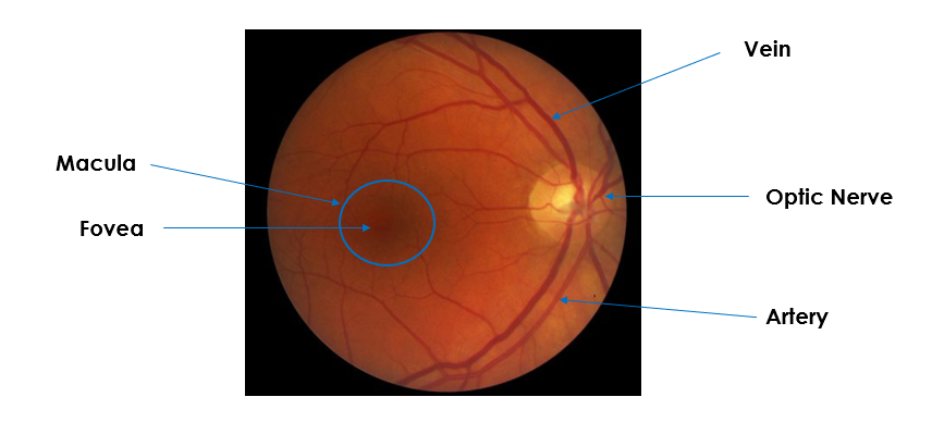
- Identify key anatomical structures visible with the direct ophthalmoscope
- Learn the parts and settings of the direct ophthalmoscope
- Learn the exam technique of the direct ophthalmoscope
Understand the utility of the direct ophthalmoscope
The direct ophthalmoscope allows you to look into the back of the eye to look at the health of the retina, optic nerve, vasculature and vitreous humor. This exam produces an upright image of approximately 15 times magnification.
Instrument Parts

Light Settings
- Large/Medium/Small/Half
- The Large aperture is used for a dilated pupil after administering mydriatic drops.
- The medium aperture is the standard for a non-dilated pupil in a dark room.
- The small aperture is for a constricted pupil in a well-lit room.
- Red free
- Used to look closely at the vasculature.
- Blue
- Used to look for corneal abrasions or ulcers with fluorescein dye.
- Slit
- Used to look at contour abnormalities of the cornea, lens or retina.
- Grid
- Used to approximate the relative distance between retinal lesions.

Exam Technique
- Wash your hands.
- Introduce yourself to the patient and explain what you are going to do.
- Position the patient so that the ophthalmoscope is held directly at the level of the patient’s eye.
- Turn on the ophthalmoscope and set the light to the correct aperture.
- Dim the lights.
- Instruct the patient to focus on an object straight ahead on the wall.
- To exam the patient’s RIGHT eye, hold the ophthalmoscope in your RIGHT hand and use your RIGHT eye to look through the instrument.
- Place your left hand on the patient’s head and place your thumb on their eyebrow.
- Hold the ophthalmoscope about 6 inches from the eye and 15 degrees to the right of the patient.
- Find the red reflex.
- Move in closer, staying nasally until you see the optic nerve.
- Rotate the diopter lens until the optic nerve comes into focus.
- The farsighted eye requires more plus/green number lenses.
- The nearsighted eye requires more minus/red number lenses.
- Measure the cup to disc ratio.
- Scan slightly up, down, right and left to look at the vessels.
- Move out temporally to find the macula and fovea.
- Repeat the same technique on the other eye.
References:
- “Examination of the optic nerve at the slit-lamp biomicroscope with a handheld lens – EyeWiki.” Accessed July 17, 2018. http://eyewiki.aao.org/Examination_of_the_optic_nerve_at_the_slit-lamp_biomicroscope_with_a_handheld_lens
- “3.5V Standard Ophthalmoscope.” Accessed July 17, 2018. https://www.welchallyn.com/en/products/categories/physical-exam/eye-exam/ophthalmoscopes–traditional-direct/35v_standard_ophthalmoscope.html
Faculty Approval by: Griffin Jardine, MD
Copyright statement: Copyright Tania Padilla Conde, ©2018. For further information regarding the rights to this collection, please visit: URL to copyright information page on Moran CORE
Identifier: Moran_CORE_25265
Disclosure (Financial or other): None